このマイクロバイブレーションがはじめて研究されたのはドイツということになっている。その後、研究は日本に移って稲垣医博が長いこと研究されて基礎を作られた。
The first stage of this Micro Vibration study was started allegedly in Germany, which later shifted to Japan under supervision of Dr. Inagaki who built the basics of this vibration.
過去の研究によれば、人のマイクロバイブレーションは、大まかに分類して3つの種類があることになっている。
1つ目は、心臓の鼓動によって発生するもの、
2つ目は、組織が疲労したり、損傷を受けた時にその部位に局部的に発生するもの、
3つ目は、通常は雑音として切り捨て処理されるもの、
の3種類がそれである。
As far the studies made in the days, micro vibrations detected on human skin can be categorized to three kinds.
These three are;
for the first, ones given by pulse,
second, ones seen on a specific area where the organs were exhausted, or a point which got any physical damage,
third, ones which are regarded as noise thus most always to be cut off.
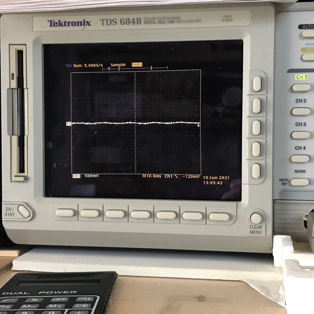
この1つ目を専門的に「心弾動図性マイクロバイブレーション」などと言ったりする。心弾動図とは動脈血管のふくらみの変化を描いたグラフである。
This first ones said above are called “Ballistocardiographic Micro vibration”. Ballistocardiograph is a graph showing the swelling movements of artery.
心臓から押し出された血液が血管を通るときは、血管壁が実際に膨らんだりしぼんだりする。心弾動図はその変化を捉えてグラフにして観察したりする。
だから心弾動図は血管の物理的動きを表しているし、もちろん原因となっている鼓動の様子も表すことになる。When blood goes through artery after being pumped out of the heart, it makes the artery wall swelled or shrinked. We use ballistocardiograph to see these movements. In other words ballistocardiograph tells the physical movements of artery, accompanied with the data given from the pulse by heart.
心電図は鼓動を生み出すための電気信号を表すから、この点が心弾動図とは異なる。On the other hand an Electrocardiograph tells the electric signals which cause pulses, not the physical movements.
日本生体マイクロバイブレーション診断技術学会(英名:Japan Society of Bio-Microbivrative Diagnostics and Technology)は、哺乳動物の生体表面に表出するマイクロバイブレーションを解析して、その哺乳動物の健康状態を診断するための検査技術に関わる研究を対象として、研究者間交流、情報交換を行うことを目的として設立される予定の、日本で唯一の生体マイクロバイブレーションを利用する診断と技術に関わる学会です。